

Vitreo Retina & Uvea Services
What is it ?
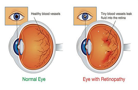
Diabetic retinopathy is a diabetic related complication that affects eye sight. It's caused by damage to the blood vessels of the light-sensitive tissue at the back of the eye (Retina). The condition can develop in anyone who has type 1 or type 2 diabetes. If left untreated, diabetic retinopathy can cause blindness.Knowing more about Diabetic Retinopathy
Classification
The diagnosis of DR is made by clinical manifestations of vascular abnormalities in the retina. Clinically, DR is divided into two stages: non-proliferative diabetic retinopathy (NPDR) and proliferative diabetic retinopathy (PDR). NPDR represents the early stage of DR, wherein increased vascular permeability and capillary occlusion are two main observations in the retinal vasculature. PDR, a more advanced stage of DR, is characterized by neovascularization. During this stage, the patients may experience severe vision impairment.What are the risk factors for DR?
 Old Age
 Genetic Factor
 Smoking/Alcohol Use
 Other diseases
 Not getting enough exercise
Vision Loss due to DME
 The most common cause of vision loss in patients with DR is diabetic macular edema (DME).
People with type 1 or type 2 diabetes can develop DME. DME occurs when excess fluid starts to build up in the macula of the eye. The macula allows us to focus and see fine details. It’s located in the center of the retina. When excess fluid builds up in the macula, it causes vision problems.DME generally develops over time. High blood sugar levels can damage the blood vessels in the retina.
Signs and Symptoms
 Spots or dark strings floating in your vision (floaters)
 Blurred vision
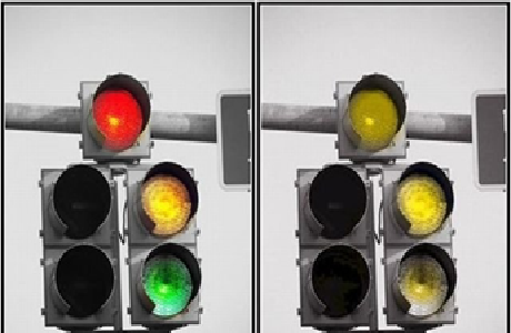 Impaired color vision
When to see a doctor
If you have diabetes, see your eye doctor for a yearly eye exam with dilation — even if your vision seems fine to prevent vision loss. Pregnancy may worsen diabetic retinopathy your eye doctor may recommend eye exams throughout your pregnancy.Treating DME
- Injectable medications.( anti VEGF and steroids)
- Laser therapy.
- Glycemic control
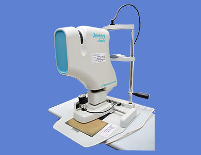
Optical Coherence Tomographs(OCT)
It is a non invasive imaging modality which uses light waves to takes optical cross sections of the light sensitive layer of the eye which is retina. Each of the ten distinctive layers of the retina can be visualized with high resolution, allowing detection and measurement of any abnormality in each of these layers. This provides useful information to diagnose several important retinal conditions such as diabetic retinopathy and Age related Macular degeneration (ARMD). It is also a very useful tool to monitor benefits of treatment in these diseases. The new feature of OCT angiography also provides valuable information on the health of various retinal vasculature without the need for invasive dye injection. OCT can also provide high resolution images of the optic disc and the anterior segment, thus becoming a valuable tool in diagnosing and monitoring glaucoma.

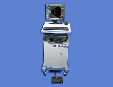
Fundus Camera & FFA
Fundus photography involves taking a picture of the light sensitive layer of the eye called the retina. The instrument consists of an intricate microscope attached to a flash enabled camera. The main structures visualized are the retina, optic disc and the macula and 60deg of the retinal periphery. It is a very useful tool to visualize and document findings in that portion of the eye which is not evident to the patient directly. It also allows monitoring the progression of the diagnosed eye conditions.
B-Scan With UBM
B-scan ultrasonography is an important adjuvant especially in opaque media for the clinical assessment of various ocular and orbital diseases. Can be used to rule out retinal, vitreous, and choroidal detachments, tumors, and other pathologies that affect the posterior segment of the eye. UBM is high resolution tool for anterior segment imaging. Indicated in opaque cornea, anterior segment trauma, hypotony, angle evaluation, IOL position.
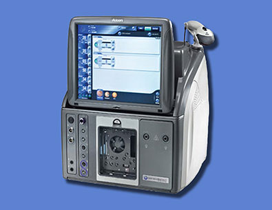
Vitrectomy-Machine
The microincision vitrectomy system (MIVS) machine is used to remove the vitreous gel from the inside of the eye which gives the surgeon better access to the back of the eye(Retina) Vitreous is a clear, jelly-like substance that fills the inside of the eye. The machine performs the basic functions of vitreous cutting with 23G/25G/27G cutters, irrigation, aspiration, and illumination. The basic components of the vitrectomy machine are viewing system, vitreous cutters, infusion system, and operating microscope. It is performed in conditions such as retinal detachment, tractional diabetic retinal detactment, macular holes, vitreous haemorrhage etc.
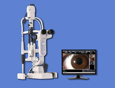
Photo Slit Lamp
Slit lamp biomicroscopy Instrument Manufacturers-TopCon. The console consists of a slit lamp biomicroscope equipped with a digital camera for capturing static and dynamic, single and multiple images of the ocular surface and anterior segment. The images are displayed and stored in the files. They can be visualized, stored, magnified and retrieved for further use. Uses:Documentation of ocular pathology with the help of clear digital photographs. Helps in counselling the patients with regards to pathology.
Get In Touch
Contact Us
Mail Us