
OCULOPLASTY SERVICES
Oculoplasty services offered at LCH-SEH: Diagnosis and Surgical Management of
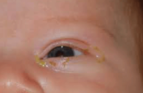
Lacrimal Diseases (Watering Eye / Tear Duct Block) in Children & Adults
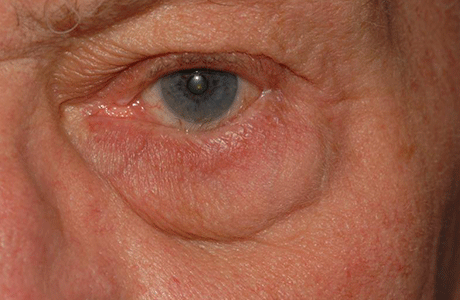
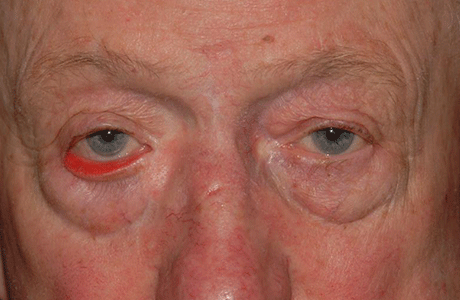
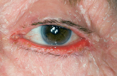
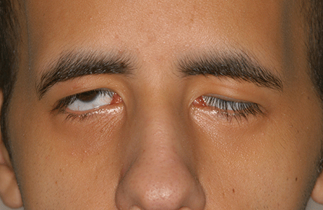
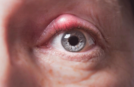
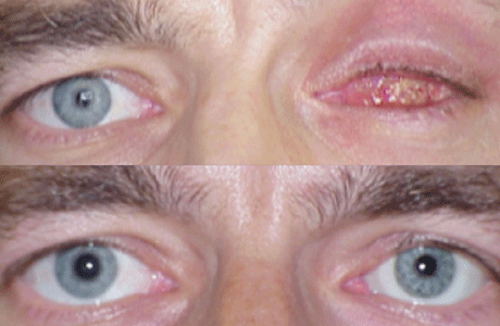
Eyelid Diseases: Entropion & Ectropion (In-Turning / Out-Turning of Eyelids)
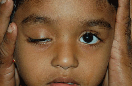
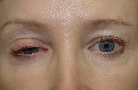
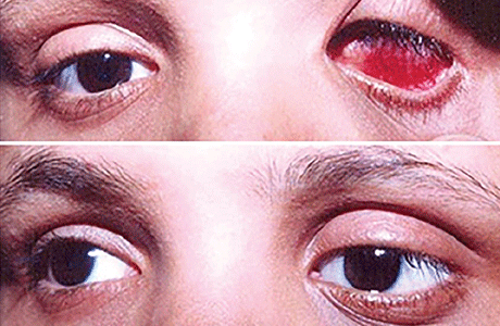
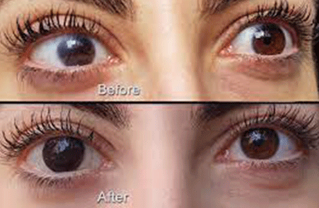
Ptosis (Droopy Eyelids)
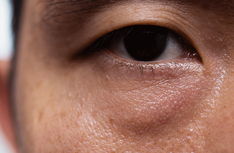
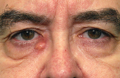
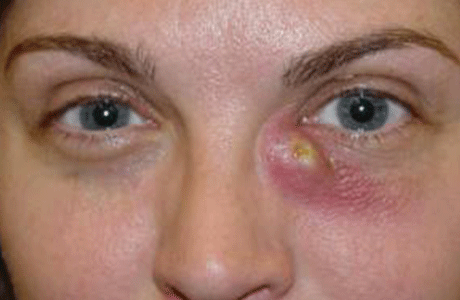
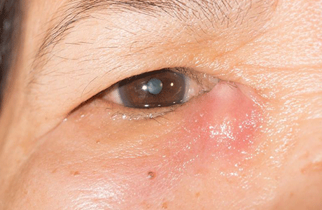
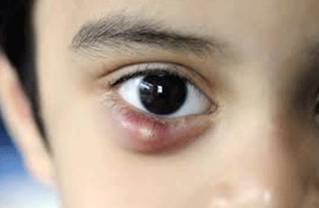
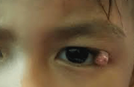
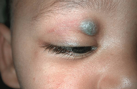
Eyelid Mass
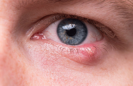
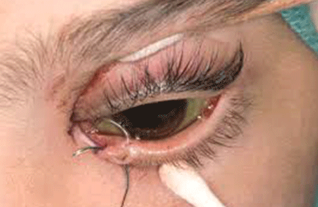
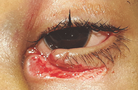
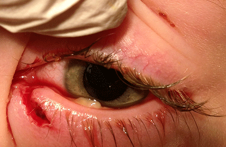
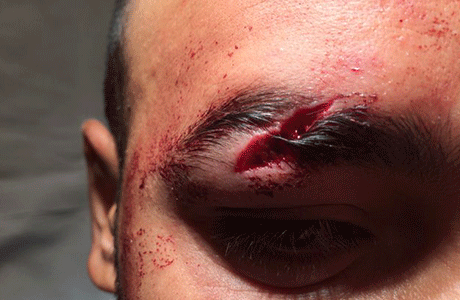
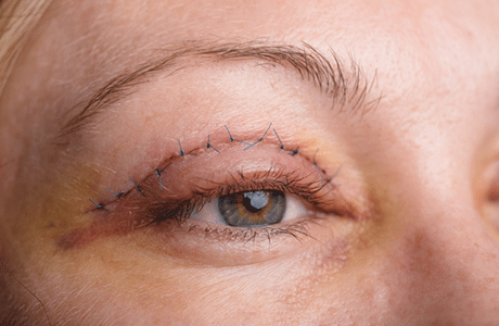
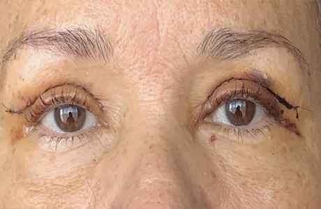
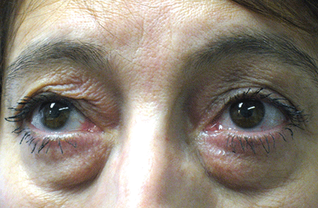
Eyelid Trauma Reconstruction
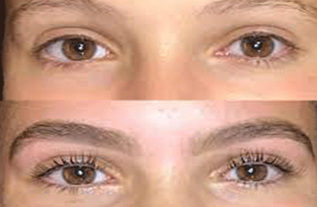
Blepharoplasty (Cosmetic Surgery for Eyelids)

Evisceration & Enucleation (Surgery for Blind / Painful Eyes)
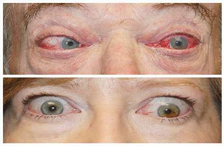
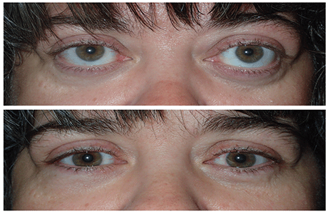
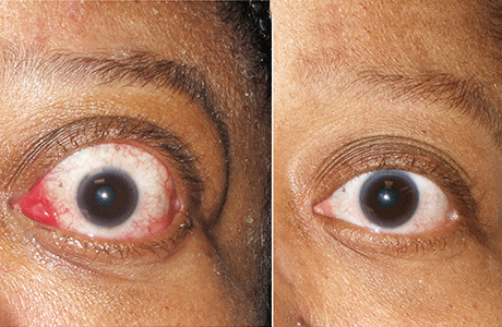
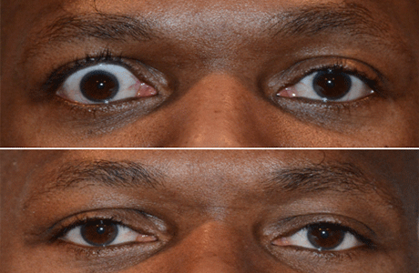
Thyroid Eye Disease
Optical Coherence Tomographs(OCT)
It is a non invasive imaging modality which uses light waves to takes optical cross sections of the light sensitive layer of the eye which is retina. Each of the ten distinctive layers of the retina can be visualized with high resolution, allowing detection and measurement of any abnormality in each of these layers. This provides useful information to diagnose several important retinal conditions such as diabetic retinopathy and Age related Macular degeneration (ARMD). It is also a very useful tool to monitor benefits of treatment in these diseases. The new feature of OCT angiography also provides valuable information on the health of various retinal vasculature without the need for invasive dye injection. OCT can also provide high resolution images of the optic disc and the anterior segment, thus becoming a valuable tool in diagnosing and monitoring glaucoma.

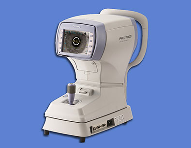
Auto Refractometer
The operator measure the refractive error and help in accurate prescription of glasses even in children and measure diameter of the cornea, pupil or hard contact lenses worn by the patient. The patient focuses on a color photo target and when measurement is taken the data displays instantly.
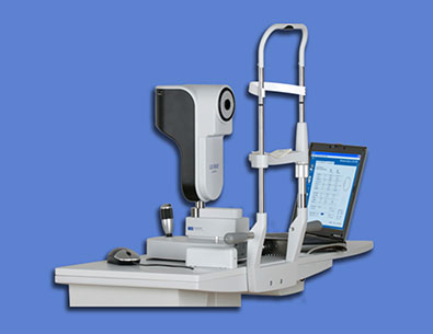
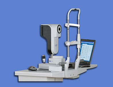
Lenstar
Lenstar is used for obtaining Ocular Measurement and performing calculations to assist in the determination of the appropriate power and type of IOL (Intra Ocular Lens) for implantation after removal of Cataract.
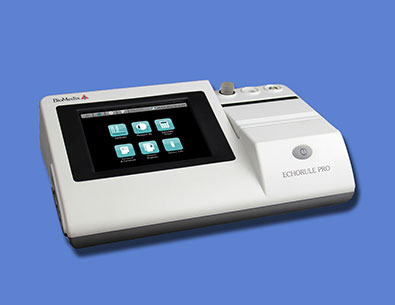
A-Scan
A-Scan is a routine type of diagnostic test used in ophthalmology. This equipment provides data on the length of the eye, which is a major determinant in common sight disorders. The most common use is to determine eye length for calculation of intraocular lens power.
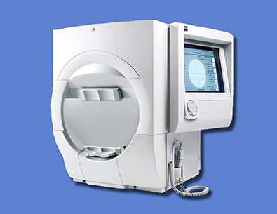
Humphery Fields Analyser
It is the newest and most advanced visual field testing platform for glaucoma and optic nerve diseases. This equipment determines the stage of disease, rate of progression and a patient's risk of future vision loss by automatically summarizing all available visual field test results, by calculating each patient's rate of visual field deterioration. With this advanced perimeter, you can more closely monitor changes in the eye to prevent irreversible loss of vision for a disease that often has no warning signs.
Yag Laser
Yag laser equipment produces short-pulsed, high-energy light beams to cut, perforate, or fragment tissue useful to improve vision when posterior capsule thickens after surgery. The YAG laser is commonly used to vaporize a portion of the capsule, allowing light to pass through to the retina. The procedure is completely painless, takes only a few minutes, and is effective in eliminating the cloudy condition.
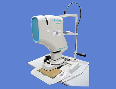
Fundus Camera & FFA
Fundus photography involves taking a picture of the light sensitive layer of the eye called the retina. The instrument consists of an intricate microscope attached to a flash enabled camera. The main structures visualized are the retina, optic disc and the macula and 60deg of the retinal periphery. It is a very useful tool to visualize and document findings in that portion of the eye which is not evident to the patient directly. It also allows monitoring the progression of the diagnosed eye conditions.
B-Scan With UBM
B-scan ultrasonography is an important adjuvant especially in opaque media for the clinical assessment of various ocular and orbital diseases. Can be used to rule out retinal, vitreous, and choroidal detachments, tumors, and other pathologies that affect the posterior segment of the eye. UBM is high resolution tool for anterior segment imaging. Indicated in opaque cornea, anterior segment trauma, hypotony, angle evaluation, IOL position.
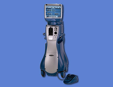
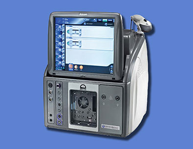
Infinity Vision System
INFINITY VISION SYSTEM is high end Phaco Machine and is designed for smoother cutting of Cataract and excellent efficiency for Micro Incision Cataract Surgery.
Vitrectomy-Machine
The microincision vitrectomy system (MIVS) machine is used to remove the vitreous gel from the inside of the eye which gives the surgeon better access to the back of the eye(retina) Vitreous is a clear, jelly-like substance that fills the inside of the eye. The machine performs the basic functions of vitreous cutting with 23G/25G/27G cutters, irrigation, aspiration, and illumination. The basic components of the vitrectomy machine are viewing system, vitreous cutters, infusion system, and operating microscope. It is performed in conditions such as retinal detachment, tractional diabetic retinal detactment, macular holes, vitreous haemorrhage etc.
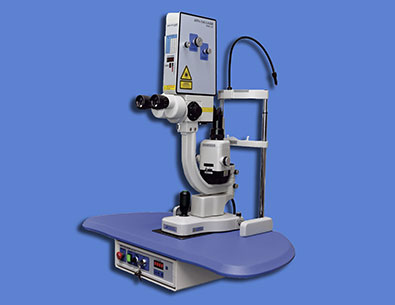
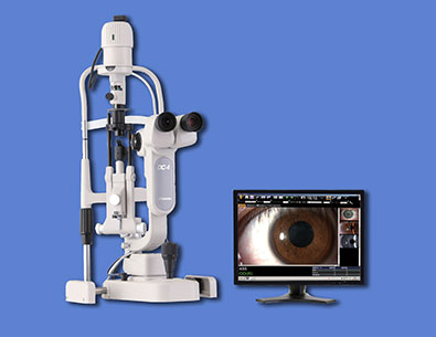
Photo Slit Lamp
Slit lamp biomicroscopy Instrument Manufacturers-TopCon. The console consists of a slit lamp biomicroscope equipped with a digital camera for capturing static and dynamic, single and multiple images of the ocular surface and anterior segment. The images are displayed and stored in the files. They can be visualized, stored, magnified and retrieved for further use. Uses:Documentation of ocular pathology with the help of clear digital photographs. Helps in counselling the patients with regards to pathology.
Green Laser Unit
This double-frequency Nd-YAG laser (532nm) is a gold-standard for treatment of many pathologies by Photocoagulation. LASER stands for Light Amplification by Stimulated Emission of Radiation. LASERs used in the OPD (Out-patient department) are mainly used for the management of retinal conditions. These are done usually to the areas of ischemia in retina especially when abnormal new vessels are formed or to weak areas in retina (holes, tears). Mode od delivery is through a Slit-lamp, through the Laser Indirect Ophthalmoscope (LIO).
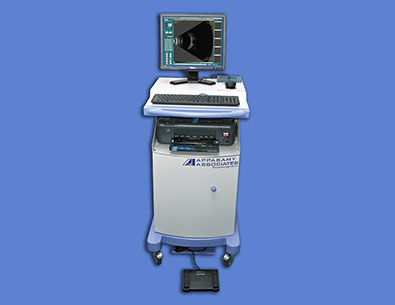
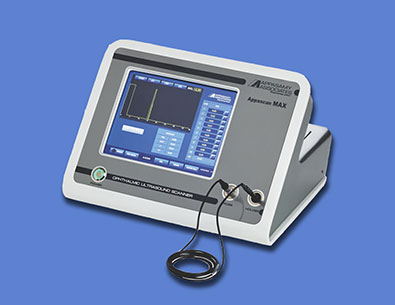
Pachymetry
Ultrasound Pachymetry Instrument: Ophthalmic Ultrasound Scanner. Manufacturers:Appasamy Associates. It is used for corneal thickness measurement by Immersion /contact method within a range of 200 to 1000 microns with an accuracy in the range of +/_5 microns. Probe frequency 10 MHz Other uses:Measurement of axial length,anterior chamber depth, Lens thickness.
Get In Touch
Contact Us
Mail Us