GLAUCOMA SERVICES
What is Glaucoma?
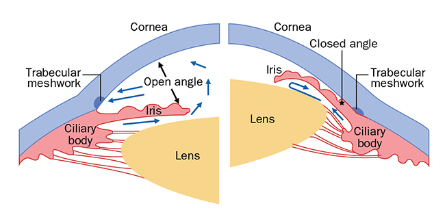
Glaucoma is a group of eye conditions that damages the optic nerve. This damage is often caused by an abnormally high pressure in your eye. The pressure increases in the eye if the passages that normally allow fluid within your eye (aqueous humor) to drain, become clogged or blocked. Fluid within your eyes then builds up and increased pressure on the optic nerve which gets damaged, resulting in vision loss. Based on the GONIOSCOPY, glaucoma is divided into “open angle” and “angle closure” Glaucoma.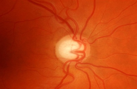
What are the symptoms of Glaucoma?
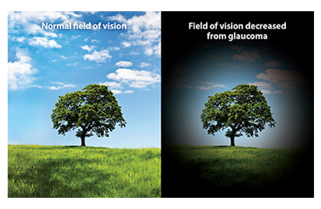 Glaucoma is called the “SNEAK THIEF OF SIGHT” as for most people, there are usually no symptoms. The first sign of glaucoma is often the loss of side/peripheral vision, which can go unnoticed until over 40% of the vision has been host.
You could be at risk of Glaucoma if you have
 High Eye pressure
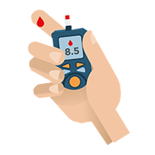 Diabetes
 Aging
Aging
 Family history of Glaucoma
 Previous Eye Injury
 High Myopia
 Steroid Medication
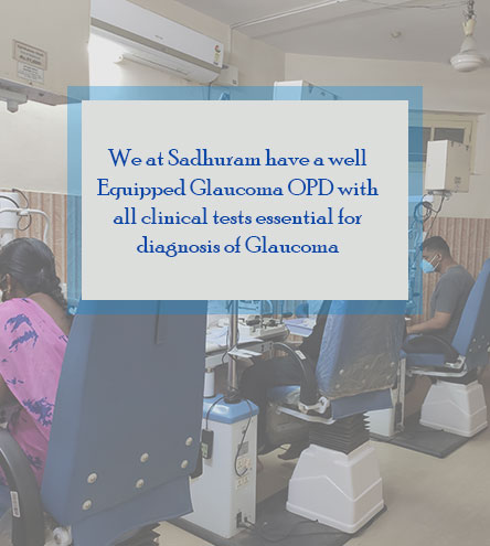
Can Glaucoma be prevented?
Early detection and treatment can control glaucoma and reduce the chances of vision loss. Any vision lost as a result of glaucoma usually cannot be restored. This is why regular preventive eye examinations are very important.What are the clinical Test Required?
1. Tonometry - Measurement of Eye pressure.2. Gonioscopy – Viewing of drainage angle .
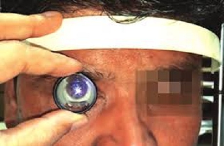
3. Disc Evaluation – Viewing of the optic nerve head at the Slit Lamp with special lens 90D, 78D.
Are they any Investigations to be done?
1. Visual field test (perimetry) to check for peripheral vision.2. Pachymetry - Thickness of the Cornea; where Eye pressure is measured.
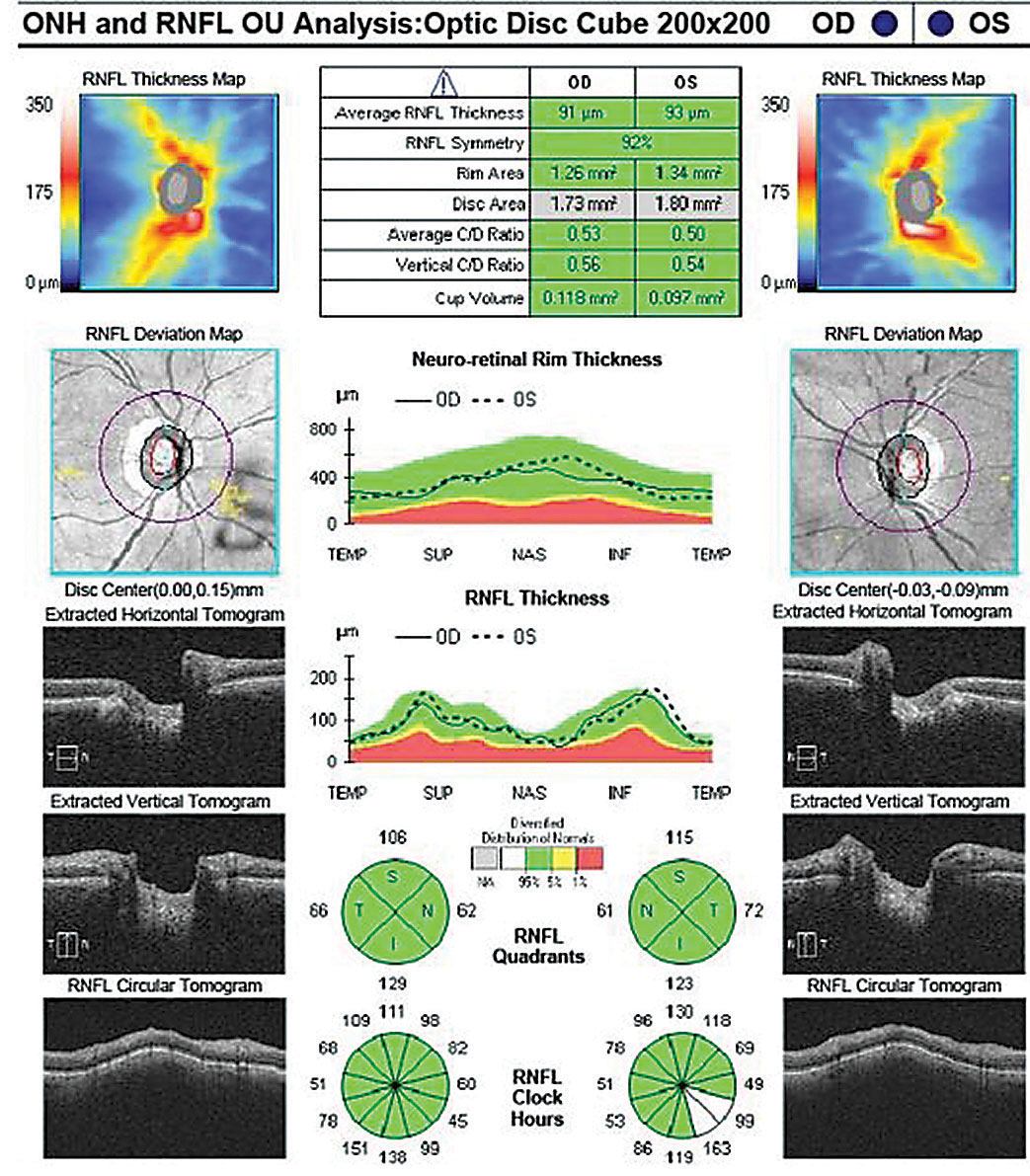
3. OCT – RNFL & ONH when suspicion of Early disease.
4. UBM – FOR ANGLE CLOSURE.
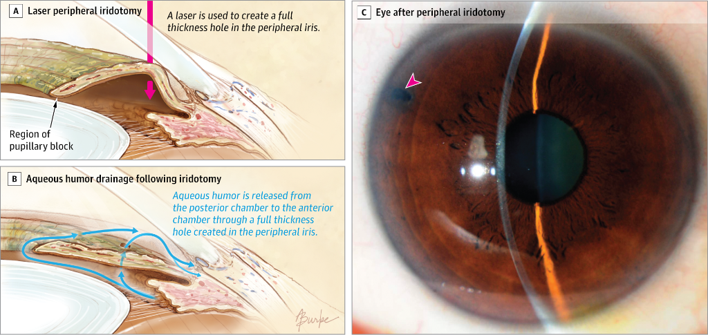
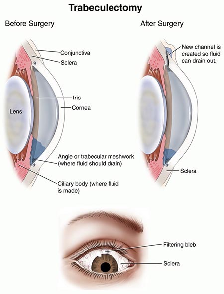
How is Glaucoma Treated?
Glaucoma is usually effectively treated with prescription of eye drops that must be taken regularly. Some cases require laser therapy or surgery. The goal of the treatment is to prevent vision loss by lowering the pressure in the eye.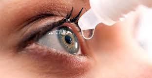

Optical Coherence Tomographs(OCT)
It is a non invasive imaging modality which uses light waves to takes optical cross sections of the light sensitive layer of the eye which is retina. Each of the ten distinctive layers of the retina can be visualized with high resolution, allowing detection and measurement of any abnormality in each of these layers. This provides useful information to diagnose several important retinal conditions such as diabetic retinopathy and Age related Macular degeneration (ARMD). It is also a very useful tool to monitor benefits of treatment in these diseases. The new feature of OCT angiography also provides valuable information on the health of various retinal vasculature without the need for invasive dye injection. OCT can also provide high resolution images of the optic disc and the anterior segment, thus becoming a valuable tool in diagnosing and monitoring glaucoma.
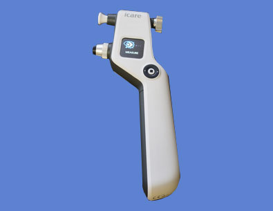
Eye Care Tonometry
Tonometry is a diagnostic test that measures the pressure inside your eye, which is called intraocular pressure (IOP). The iCare Tonometer provides accurate, quick and painless measurement of the intraocular pressure. This device uses a rebound measuring principle which requires no drops, no puff of air.
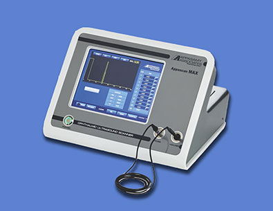
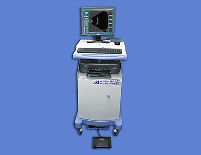
Pachymetry
Ultrasound Pachymetry Instrument: Ophthalmic Ultrasound Scanner. Manufacturers:Appasamy Associates. It is used for corneal thickness measurement by Immersion /contact method within a range of 200 to 1000 microns with an accuracy in the range of +/_5 microns. Probe frequency 10 MHz Other uses:Measurement of axial length,anterior chamber depth, Lens thickness.
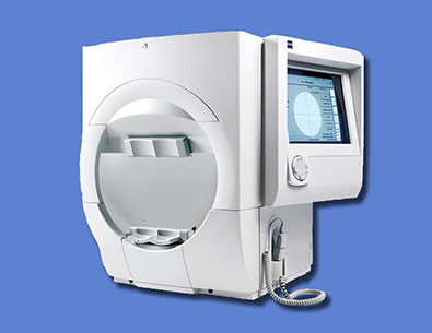
Humphery Fields Analyser
It is the newest and most advanced visual field testing platform for glaucoma and optic nerve diseases. This equipment determines the stage of disease, rate of progression and a patient's risk of future vision loss by automatically summarizing all available visual field test results, by calculating each patient's rate of visual field deterioration. With this advanced perimeter, you can more closely monitor changes in the eye to prevent irreversible loss of vision for a disease that often has no warning signs.
B-Scan With UBM
B-scan ultrasonography is an important adjuvant especially in opaque media for the clinical assessment of various ocular and orbital diseases. Can be used to rule out retinal, vitreous, and choroidal detachments, tumors, and other pathologies that affect the posterior segment of the eye. UBM is high resolution tool for anterior segment imaging. Indicated in opaque cornea, anterior segment trauma, hypotony, angle evaluation, IOL position.
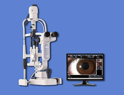
Photo Slit Lamp
Slit lamp biomicroscopy Instrument Manufacturers-TopCon. The console consists of a slit lamp biomicroscope equipped with a digital camera for capturing static and dynamic, single and multiple images of the ocular surface and anterior segment. The images are displayed and stored in the files. They can be visualized, stored, magnified and retrieved for further use. Uses:Documentation of ocular pathology with the help of clear digital photographs. Helps in counselling the patients with regards to pathology.
Get In Touch
Contact Us
Mail Us